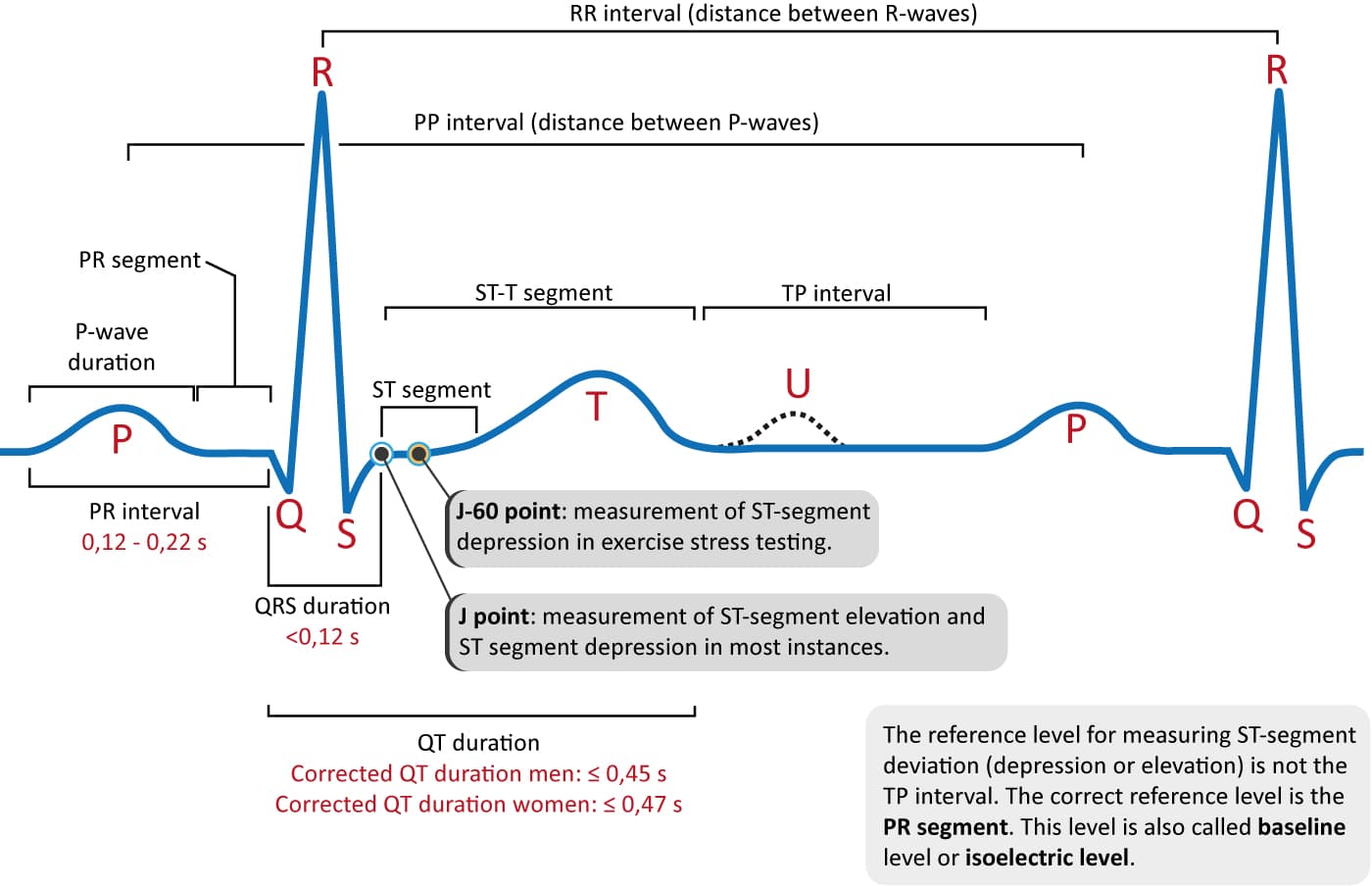
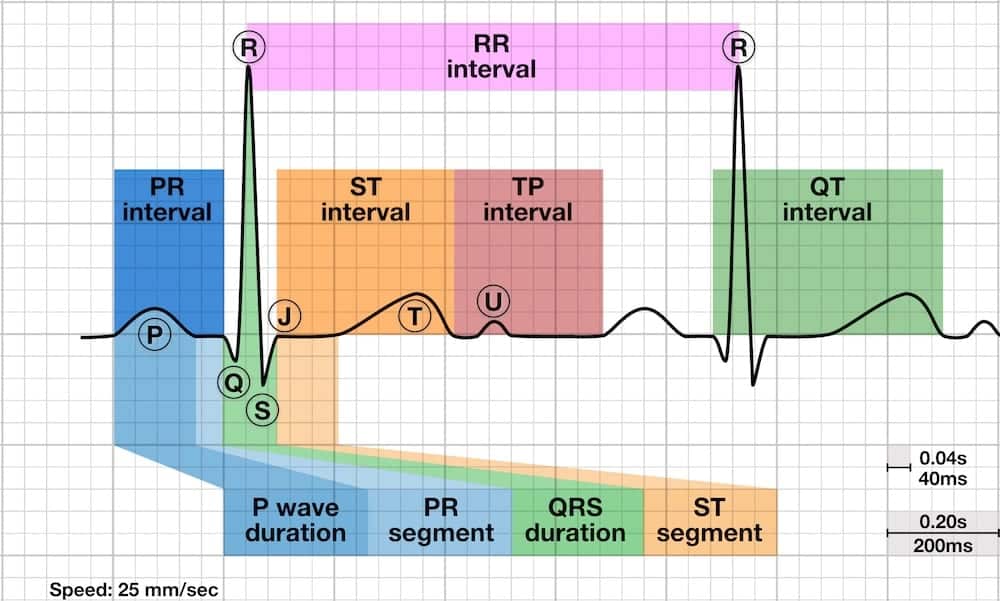
The Deflection Waves in an Ecg Tracing Include
High takeoff of r that not necessary coincides with J point 2 mm. The information needed include.

Pin On All About Nursing Things To Help Me Remember Because My Brain Is So Full That It May Explode
Filling the forms involves giving instructions to your assignment.

. A positive or negative deflection from baseline that indicates a specific electrical event. The typical characteristics of Type2 Brugada ECG pattern or saddleback pattern in leads V 1 V 2 include the following Figs. 6 and and12 12.
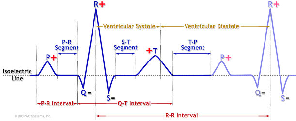
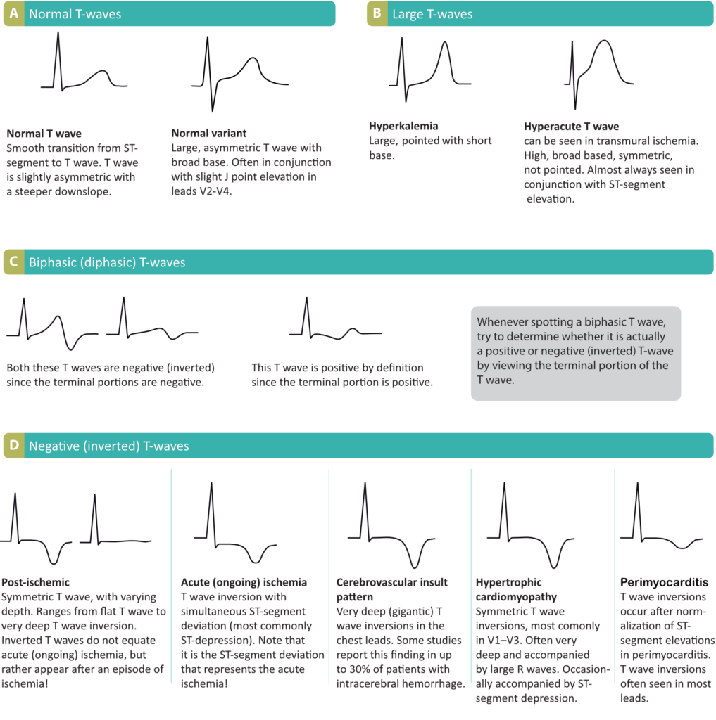
Click on the order now tab. You will be directed to another page. The waves on an ECG include the P wave Q wave R wave S wave T wave and U wave.
Here there is a form to fill. It is very easy. Topic subject area number of pages spacing urgency academic level number of sources style and preferred language style.
Minimum STsegment ascent. Descending arm of r coincides with the beginning of ST sometimes is not well seen.

T Waves In Normal Ecg Signal A Possible T Waves Forms In Download Scientific Diagram
Chapter 18 Cardio Physiology Dr Parker Pharmacology Nursing Medical School Essentials Nursing School Studying
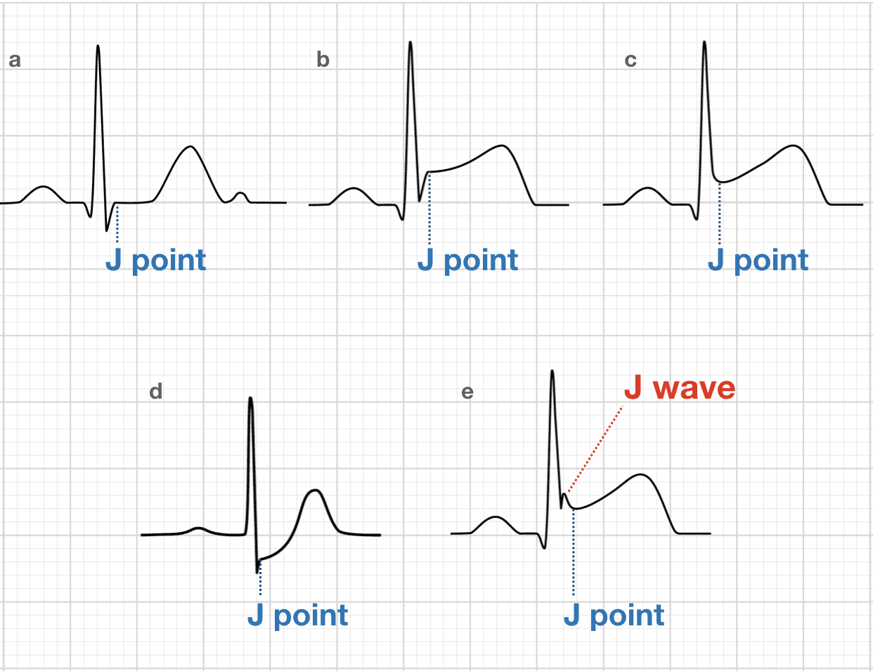
J Point Ecg Interval Litfl Ecg Library Basics

Ecg Interpretation Characteristics Of The Normal Ecg P Wave Qrs Complex St Segment T Wave Ecg Echo
Dr Smith S Ecg Blog March 2022

Different Types Of P Wave Morphology Typical Examples Of Type 1 P Download Scientific Diagram

How To Read An Ekg Electrocardiograph Pr Interval Medical Library Ekg Interpretation

Electrocardiographic T Wave Morphology And Risk Of Mortality International Journal Of Cardiology

What Is An Ecg
Ecg Interpretation Characteristics Of The Normal Ecg P Wave Qrs Complex St Segment T Wave Ecg Echo

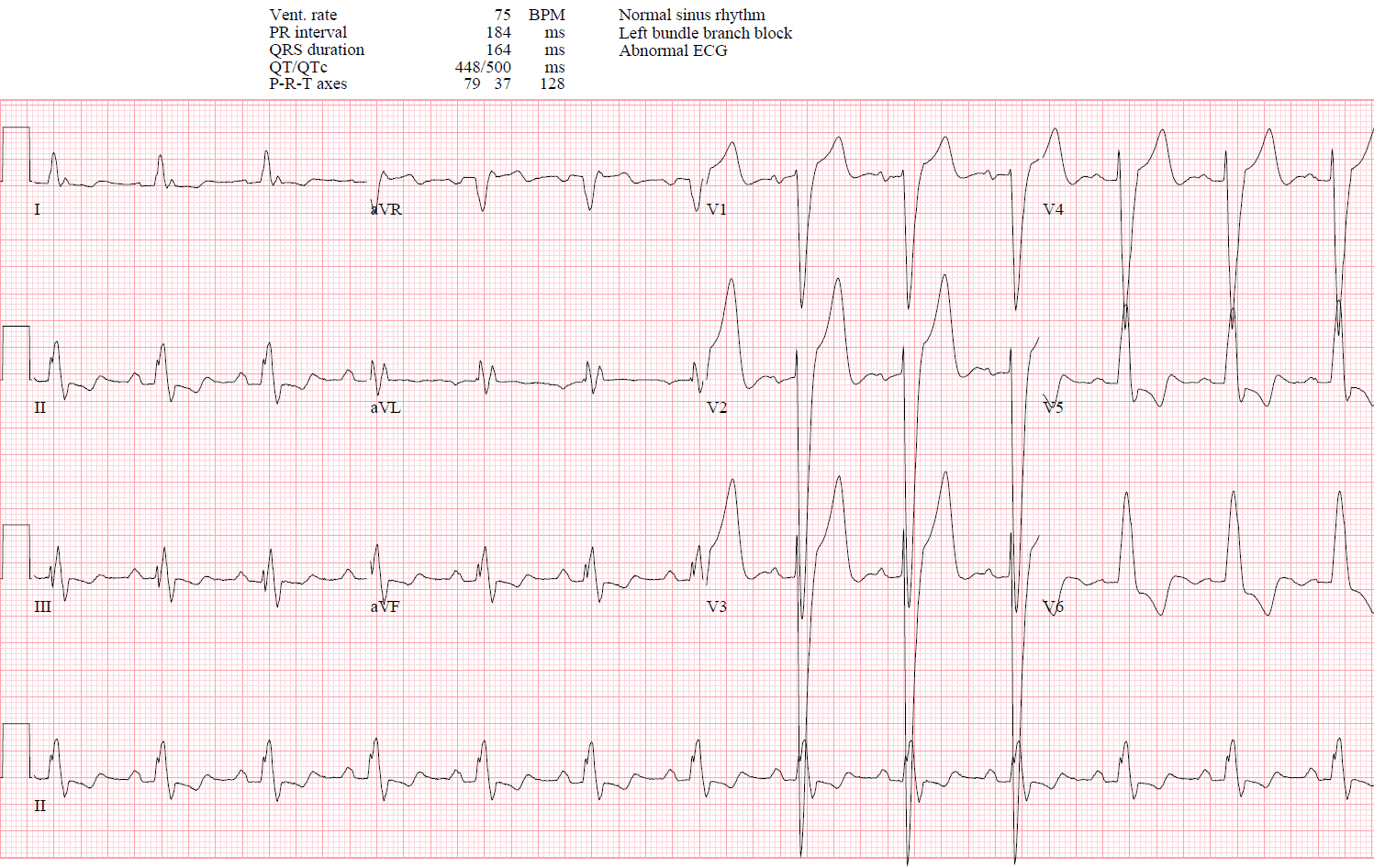
Right Bundle Branch Block Qrs 0 12 S Terminal R Wave In Lead V 1 Download Scientific Diagram
Qrs Amplitiude Respiratory Modulation Biopac
Ecg Interpretation Characteristics Of The Normal Ecg P Wave Qrs Complex St Segment T Wave Ecg Echo
J Point Ecg Interval Litfl Ecg Library Basics

Ecg A Pictorial Primer

Illustration Of Global Qrs Onset And Local T Wave Landmarks Detection Download Scientific Diagram
Ecg Interpretation Characteristics Of The Normal Ecg P Wave Qrs Complex St Segment T Wave Ecg Echo

Ecg A Pictorial Primer
Epsilon Wave Small Negative Deflection Just Beyond The Qrs In V 1 Or V 2 Download Scientific Diagram


Comments
Post a Comment